The elearning for healthcare (elfh) Clinical Imaging programme has been developed by the Society and College of Radiographers (SCoR) in partnership with NHSE.
Over 16 years it has transformed healthcare education increasing in scope and depth to provide a comprehensive resource which is CPD Now endorsed by the College of Radiographers and is valued by radiographers and healthcare professionals in the UK.
It’s great to see that our partnership with eIntegrity has enabled this resource to be accessed internationally and Clinical Imaging is now available in 140+ countries across five continents.
Medical Imaging (Clinical Imaging)
Develop your understanding of clinical imaging – how to interpret x‑ray, CT, MRI and ultrasound images, and the roles and responsibilities of professionals delivering safe, effective diagnostic imaging. Includes targeted learning on anatomy, pathology, cross‑sectional techniques and real‑case interpretation to support confident clinical decision‑making.
Multiple online learning modules
Engaging multimedia‑based learning for anyone involved in clinical imaging
Who is this Medical Imaging course for?
- Radiographers
- Sonographers
- Clinical imaging staff
- Medical imaging trainees
- Forensic imaging practitioners
- CT and MRI practitioners
- Nuclear medicine staff
- Emergency imaging teams
- Paediatric imaging practitioners
What’s on this page?
Introduction to Medical Imaging
This learning provides a comprehensive introduction to clinical imaging, helping you develop the skills needed to interpret radiological images accurately and confidently. It covers essential anatomy, pathology, imaging technologies and the principles that underpin safe, effective diagnostic practice across a wide range of clinical settings. You’ll explore everything from plain film x‑ray interpretation to cross‑sectional imaging, including CT, MRI, ultrasound and Doppler scanning, along with modules on paediatric imaging, forensic radiography, musculoskeletal imaging and more.
Developed in the UK by the College of Radiographers and NHS England’s elearning for healthcare programme, this course reflects high standards of radiographic practice and clinical imaging provision. Highly interactive sessions, real patient cases, animations and quizzes help you build strong interpretation skills and expand your understanding of both normal variants and complex pathologies.
After completing this course, you should be able to:
- Describe the fundamentals of diagnostic imaging and how different modalities are used
- Identify normal anatomy and distinguish anomalies, fractures, suspected injuries and a wide range of pathologies across appendicular and axial skeleton systems
- Interpret CT, MRI, ultrasound and Doppler clinical images confidently
- Recognise key imaging appearances across MSK, gastro‑intestinal, genito‑urinary, obstetric, gynaecological and paediatric imaging
- Understand mechanisms of injury, including ballistic and gunshot trauma, and apply the principles of forensic radiography
- Apply safe imaging protocols, professional standards and governance responsibilities in clinical practice
You can also:
- Strengthen your communication and interpretation skills using real clinical data
- Build confidence in evaluating subtle normal variants, non‑accidental injury indicators and complex radiographic appearances
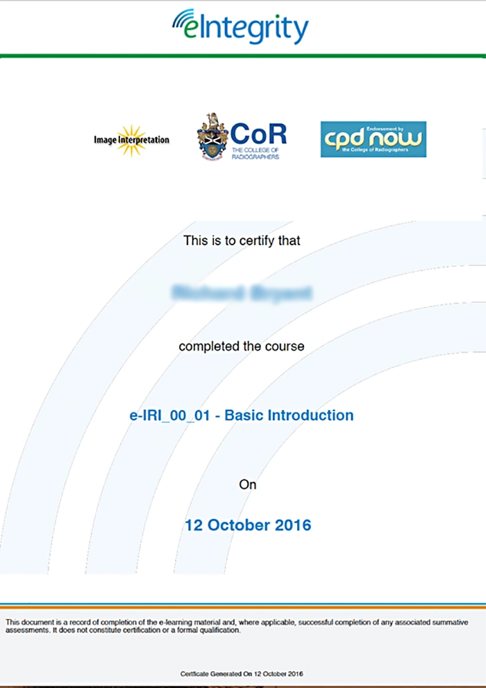
- Support your clinical development with formally endorsed CPD content

Course features
- A comprehensive radiographic and clinical imaging programme covering anatomy, pathology, technology and interpretation across multiple modalities.
- Highly interactive content featuring real clinical images, videos, animations and questions that help develop confident interpretation skills.
- Practical, case‑based and scenario‑based learning suitable for radiographers, ultrasonographers and other imaging professionals around the world.
Why choose eIntegrity?
Access high‑quality clinical imaging training developed by leading UK radiography specialists, with content aligned to recognised professional standards. This course gives mixed‑role teams a clear, structured understanding of imaging interpretation, enabling faster decisions about suitability and fit for clinical practice.
Share a simple overview with radiology departments, imaging leads or training supervisors to help them assess how the programme supports diagnostic roles across emergency, forensic, paediatric and general clinical settings.
Clear signposting throughout helps you distinguish what is core national guidance from what is shaped by local imaging protocols, service models or technology provision.
Course content overview
This programme‑level learning focuses on real‑world clinical image interpretation and the essential knowledge needed for safe diagnostic decision‑making. You’ll explore anatomy, pathology and imaging technique across x‑ray, computed tomography, magnetic resonance imaging, ultrasound and nuclear imaging modalities. Sessions cover clinical governance, patient care, consent, communication and the professional responsibilities involved in imaging practice.
Advanced modules explore neuroradiology, cardiac imaging, molecular imaging, vertebral assessment, fracture detection and mechanisms of injury. Forensic radiography content covers coroner‑related imaging, ballistics, bullets, gunshot wounds, trauma mechanism assessment and non‑accidental injury indicators.
Subject areas covered:
Anatomy, pathology, musculoskeletal imaging, fracture interpretation, cross‑sectional imaging, CT scanning, MRI resonance imaging, ultrasound and Doppler, obstetric and gynaecological imaging, paediatric imaging, forensic radiography, molecular imaging, normal variants, GI and GU imaging, cardiac imaging, vertebral assessment, quality improvement and clinical governance.
What’s included:
Highly interactive sessions with images, animations, videos, self‑assessment questions and real case material, arranged into short modules to support flexible, blended learning.
Who developed this course?
This course has been developed by the College of Radiographers in partnership with NHS England’s elearning for healthcare programme. Written by experienced clinicians and subject‑matter experts, it reflects the highest standards of radiographic practice, clinical imaging technology and diagnostic interpretation.
Our Medical Imaging modules
- Plain film x‑ray interpretation
- Anatomy and normal variants
- Musculoskeletal and fracture imaging
- Cross‑sectional technologies (CT and MRI)
- Ultrasound and Doppler
- Gastro‑intestinal imaging
- Genito‑urinary imaging
- Obstetric and gynaecological imaging
- Paediatric imaging
- Forensic radiography and ballistics
- Cardiac and molecular imaging
- Dental imaging
- Neuroradiology
- Nuclear imaging and gamma technologies
- Pathology and biochemistry in imaging
- Quality improvement and clinical governance

Access 24/7
The Medical Imaging course is delivered entirely online, giving you the flexibility to study at home, at work or on the move. Short, focused sessions make it easy to maintain progress while managing clinical shifts or study commitments.
With over 45 modules, you can dip in and out whenever you need to revisit anatomy, refine image interpretation skills or explore advanced imaging modalities. All materials remain available for reference and ongoing development.
While the programme is formally endorsed for CPD, you can also record completed sessions in your professional portfolio to support local training and competency requirements.
People also ask
Who is this course designed for?
How long does each session take?
What imaging modalities are covered?
Can the course support CPD?
Does the programme include real clinical cases?
FAQs
How can I purchase a licence to access the course?
Can I buy a licence for more than one course?
Do I have to complete the training all at once?
Will I receive a formal qualification on completion?
How do I access the content once I have paid?
Reviews & Testimonials
Comparing the cost of Medical Imaging Training
Traditional imaging training often relies on face‑to‑face workshops, case‑based tutorials or supervised reporting sessions, which can be difficult to schedule alongside clinical duties. With this online Medical Imaging course, you gain a full suite of structured modules for one straightforward subscription fee, making it a more predictable and cost‑effective option than arranging multiple standalone training sessions.
How to get started
Simply enrol through the eIntegrity platform and begin working through the modules at your own pace. Each session is designed to help you build confidence in anatomy, pathology, modality‑specific interpretation and imaging protocol without interrupting your clinical responsibilities.
Who is this programme suitable for?
This course is designed for professionals involved in delivering or supporting clinical imaging services, including:
- Radiographers
- Sonographers
- Clinical imaging practitioners
- CT and MRI staff
- Nuclear medicine teams
- Forensic imaging practitioners
- Emergency imaging staff
- Paediatric imaging specialists
- Those working in reporting or interpretation roles
The programme supports anyone looking to strengthen their understanding of radiographic practice, build confidence in anatomy and pathology interpretation and apply structured, safe imaging principles across modalities including x‑ray, CT, MRI, ultrasound and nuclear imaging.