Course Features
- Comprehensive coverage of all aspects of radiological medical imaging
- Highly interactive content, with images, videos, animations and questions
- Test your interpretation skills using patient case studies
£105.00 Excludes VAT where applicable.
This course is available on a 12-month payment plan from £9.50/month.
Multiple or multi-year licences are also available, please contact us for details.
Contact UsAny questions? FAQ's
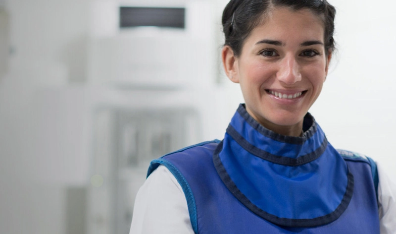
Medical Imaging (Clinical Imaging)
Our radiology clinical imaging course equips you with the skills to interpret radiological images correctly.
It is relevant to radiographers, ultrasonographers and other healthcare professionals across the world.
The programme has been developed in the UK by the College of Radiographers and NHS England elearning for healthcare.
High-Quality Online Radiography Course
You can use the sessions to develop your skills in radiography, CT, MRI and ultrasound.
There are also sessions on patient care, consent, clinical governance and professional development.
The sessions are highly interactive, with images, video clips, animations and self-assessment questions to help embed learning and understanding.
Test your interpretation skills using real clinical data in our medical imaging online course
You can view medical images from real cases and test your interpretation skills.
This helps you to better understand the wide-ranging and complex appearances of both normal radiology images and those showing pathology.
The course content is available online so you can study anywhere, at any time.
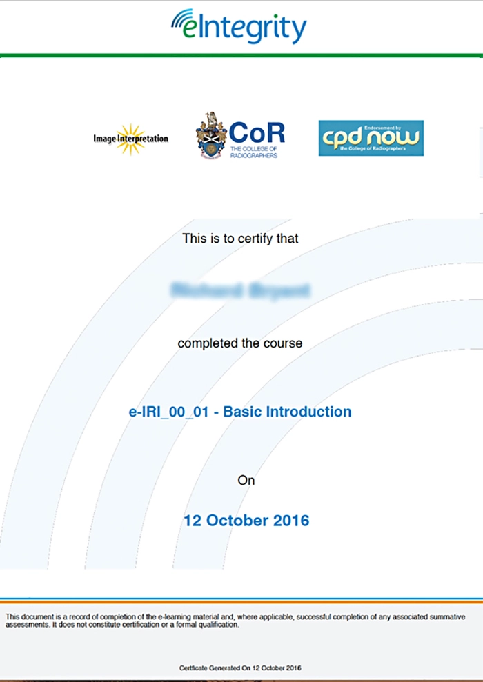
Formally Endorsed by the UK College of Radiographers
This interactive medical imaging course is formally endorsed for continuing professional development by the UK College of Radiographers (through the ‘CPD Now’ scheme). So, it meets the highest UK quality standards of a leading professional body.