Award-winning radiology training online
The Radiology – Integrated Training Initiative (R-ITI) is a high-quality e-learning resource for radiologists around the globe.
You can choose from more 750 interactive sessions that cover all aspects of radiology. The programme offers ideal preparation for FRCR* and equivalent examinations. It is also suitable for fully qualified clinicians looking to reinforce or strengthen core knowledge.
Test your diagnostic skills with interactive exercises
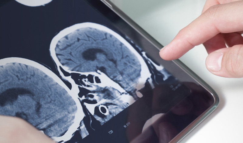
This highly engaging content includes many interactive features, such as practical exercises and self-assessment questions, to reinforce your learning and understanding. Using interactive case studies, you can analyse written reports and enhance your skills in interpreting clinical information. You can also view numerous high-quality images (including X-rays), ultrasound video clips and 3D diagrams, which creates greater spatial awareness and realism than textbook learning.
There is also a separate pathway dedicated to clinical oncology (details in the course content section).
Suitable for healthcare professionals globally
This radiology course is available online so you can study at your own pace, in your own time. R-ITI has set a gold standard for e-learning in healthcare in the UK and it is now available to healthcare professional globally.
The programme has been developed in the UK by The Royal College of Radiologists and Health Education England e-Learning for Healthcare.
*Fellowship of The Royal College of Radiologists
Watch the short course film
Course content
R-ITI features 15 modules that are divided up into smaller e-learning sessions. Each session typically takes around 30 to 40 minutes to complete.
Take a look at the course overview below or download the full R-ITI curriculum.
Clinical Oncology pathway
This standalone pathway features around 100 dedicated oncology sessions from the R-ITI programme. This pathway is suitable for all healthcare professionals working in oncology
It is included in the full R-ITI programme or available to purchase as a separate individual module. See the 'buy now' section below for details.
Module 1b – Thoracic and Respiratory
• Airway Anatomy
• CT Anatomy: Lobes, Segments and Airways
• CT Anatomy: Pulmonary Lobule and Fissures
• Staging of Lung Cancer
• Imaging of Lung Cancer
• Thoracic Lymphoma
• Diagnosis of Pulmonary Embolism
• Guidelines for the Management of Suspected Acute Pulmonary Embolism
• Malignant Mesothelioma
• Anterior Mediastinal Masses
• Mediastinal Masses: Middle and Posterior Mediastinum
• Causes of Mediastinal Nodal Disease
• Superior Vena Cava Obstruction: CT Imaging Findings of Pathology and Anatomy
%MCEPASTEBIN%
Module 1c – Vascular and Interventional
- Superior Vena Cava Obstruction: Imaging and Treatment
- Venous Interventions in the Liver: TIPS and Budd-Chiari Syndrome
Module 2 – Musculoskeletal and Trauma
- Lower Back Pain Imaging Approach
- Radiculopathy
- Benign and Malignant Lesions of the Ribs
- Sacral Lesions
- The Sclerotic Clavicle
- Approach to a Bone Lesion
- Bone Lesions of the Epiphysis
- Bone Lesions of the Diaphysis
- Mass Posterior Knee
- Spinal Tumours
- Surface Lesion of Bone
Module 3 – Gastro-intestinal
• Oesophagus: Tumours of the Oesophagus
• Oesophagus: Staging of Tumours
• Tumours of the Oesophagus: Treatments
• Neoplastic Conditions of the Stomach and Duodenum
• Malignant Tumours of the Small Bowel
• Small Bowel: G-I Stromal Tumours (Hollow Organ and Mesentery)
• Carcinoma of the Colon
• Staging Colorectal Cancer
• Colorectal Cancer: Follow-Up and Treatment, Management of Recurrence
• Imaging of the Solid Viscera
• Pharynx: Normal Anatomy and Examination Techniques
• Gallbladder and Biliary Tract: Staging of Cholangiocarcinoma
• Normal Liver Anatomy and Examination Techniques
• Liver: Malignant Tumours of the Liver – Hepatoma
• Malignant Tumours of the Liver: Others
• Pancreas: Normal Anatomy and Examination Techniques, Anomalies and Anatomic Variants
• Pancreatic Adenocarcinoma
• Other Pancreatic Neoplasms
• Staging of Pancreatic Adenocarcinoma
• Staging Stomach Cancer
%MCEPASTEBIN%
Module 4a – Breast
- Introduction to Breast Imaging
- Breast Calcification
- Staging of Breast Cancer and Metastatic Disease
Module 4b – Gynaecology-Obstretrics
- Staging Cervical and Endometrial Cancer
- Ovarian Cancer: Imaging at Diagnosis
- Gynaecological Cancer: Imaging in Follow-Up
- Complications of Treatment
Module 4c – Genito-urinary and Adrenal
- Anatomy of the Kidney
- Anatomy of the Ureter
- Anatomy of the Bladder
- Anatomy of the Prostate
- CT of the Urinary Tract
- MRI of the Urinary Tract
- TRUS Guided Biopsy of the Prostate Gland
- Benign Renal Tumours
- Renal Cell Cancer I
- Renal Cell Cancer 2
- Bladder Cancer
- Clinical Aspects, Epidemiology and Diagnosis of Prostate Cancer
- Staging Prostate Cancer
- Malignant Testicular Tumours
- Cancer of the Penis
- Imaging of Indeterminate Renal Mass
- Imaging of an Incidental Adrenal Mass
Module 5 – Paediatrics
- Hilar Enlargement
- Neuroblastoma
- Renal Tumours
- Infratentorial Tumours
- Supratentorial Tumours
- The Child Who Has Gone Off Their Legs
- Intraspinal Tumours
Module 6a – Head and Neck
- Skull Base Including the Temporal Bone
- Imaging Anatomy of the Suprahyoid Neck
- Salivary Glands
- Imaging Neck Masses in Adults
Module 6b – Neuroradiology
- Neurovascular Anatomy
- Functional Neuroanatomy
- Primary Tumours: Gliomas, Astros and Oligos
- Choroid Plexus, Glioneuronal and Pineal Region Tumours
- Medulloblastoma
- Primary Tumours: Pituitary and Hypothalamus
- Characteristic Appearances and Differential Diagnosis of Meningiomas
- Vestibular Schwannomas
- Intracranial Metastases: Imaging and Differential Diagnosis
- Central Nervous System Lymphoma
- Radiation Injury
- Primary Tumours of the Spinal Cord
- Spinal Extramedullary Tumours
- Spine Secondaries
- CT Spinal Imaging
- Petrous Temporal Bone: Infection, Cholesteatoma and Glomus Tumour
- Facial Sinuses
- Emergency CT Brain Interpretation
- Spinal Imaging
- Plain Skull Radiograph
Module 7 – Radionuclide Radiology
- Bone: Basic Concepts and Normal Bone
- Primary Tumour and Metastasis
- The Role of Nuclear Medicine in the Management of Thyroid Cancer
- Static Renal Imaging: DMSA
- Monitoring the Effects of Cancer Therapy
- Colorectal Cancer 18 FDG PET/CT
- Gastro-oesophageal Cancer 18 FDG PET/CT
- Lung Cancer FDG PET
- Lymphoma 18 FDG PET Study
- Neuroendocrine Imaging with mIBG Somatostatin Receptor Scintigraphy (SRS)
- Malignant Melanoma 99m Tc: Sentinel Node
- Breast Cancer Sentinel Node
Module 8a – Physics
- Radionuclide Imaging Overview
- Positron Emission Tomography (PET)
There is also a standalone clinical oncology pathway, which features around 100 relevant sessions from the R-ITI programme. This specific pathway is suitable for all healthcare professionals working in oncology. It is included in the full R-ITI programme or it is available to purchase as an individual module. See the 'buy now' section below for details.
For a full list of all the modules and sessions for our programmes, please go to: http://portal.eintegrity.org/catalogue
Free trial sessions
Below you can complete some sample e-learning sessions for free. See how the e-learning works and how the interactive features enhance knowledge and understanding.
Just click on the play button to view each session.
Making the most of R-ITI Trainer Guide
This introductory session to the e-learning solution, the Radiology - Integrated Training Initiative (R-ITI), is designed to give you an overview of the content, the benefits of integrating e-learning into your training design as well as a guide on how to use the materials effectively.
Making the most of R-ITI Trainee Guide
This introductory session to the e-learning solution, the Radiology - Integrated Training Initiative (R-ITI), is designed to give you an overview of the content, the benefits of learning using the R-ITI e-learning, as well as a guide illustrating how to use the materials effectively.
Bone Lesions of the Epiphysis
This session enables learners to understand the lesions that predominate in the epiphyseal area. Causes and examples of the common lesions are included.
Paediatric Bone
This session covers the skull, axial skeleton and upper limbs of paediatric bone scans.
Cardiac Tumours
In this session you will learn about the more common types of cardiac tumour. The imaging approach in these patients is discussed and the cross-sectional imaging appearances of some of these tumours is presented.
Cardiac Shapes
This session reviews the normal cardiac outline and which chambers contribute to this, followed by a review of specific chamber enlargement or absence and how this will alter the normal cardiac shape. A review of different diagnoses which result in characteristic cardiac shapes enabling the diagnosis to be made from the chest radiograph. Associated features such as other mediastinal findings and changes in the lungs will be included, where relevant e.g. right aortic arch.
Abnormalities of the Bladder and Urethra
In this session you will learn about congenital abnormalities of the bladder and urethra. You will also learn about some normal anatomical structures in the urethra which can be confused with abnormalities.
Vestibular_Schwannomas
In this session, we will cover the anatomy of the cerebellopontine angle (CPA) and the common types of CPA lesions with emphasis on vestibular schwannomas. We will briefly discuss the presentation of such lesions and the strategy for their investigation, including the role of computed tomography (CT), magnetic resonance imaging (MRI) and advanced MRI sequences.
Bone Lesions of the Diaphysis
In this session you will see a range of diaphyseal lesions. Images of aggressive and non-aggressive lesions are presented, together with explanations regarding the pathologies and their differential diagnoses.
Certificates
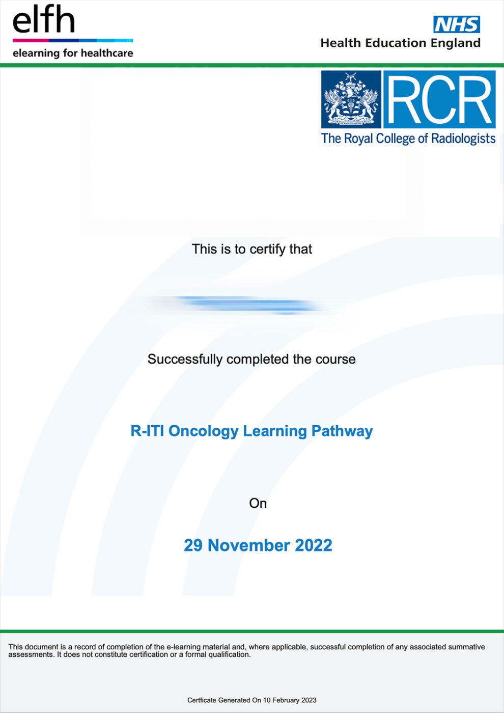
On completion, you can download and print off a certificate that is endorsed by the relevant professional body.
Buy now
Access to the full programme |
Cost |
Single-user access for 12 months |
£600* |
*excluding VAT where applicable
Access the full programme
To purchase a licence, please click on the Buy Now button below.
Multiple or multi-year licences are available. You can also pay by monthly subscription. Please contact us for details.
Buy individual modules
As an alternative, you can purchase individual modules covering specific areas and specialties, such as cardiac or paediatrics.
Just click on the relevant link(s) below. Further modules can be purchased at any time.
Prices for individual modules (excluding VAT where applicable):